

When sitting around drinking late at night, the conversation inevitably turns to superpowers and which one everyone wants.
Aside from a super healing factor and flight, one of the top picks is surely invisibility–though usually for some pretty diabolical reasons, like stealing your neighbor’s newspaper.
While we’re still not quite at the level of full-on invisibility cloaks, scientists have developed a way to make cells transparent! Invisible Woman this isn’t, but maybe it’s enough to sneak a midnight snack?
(Warning: some images may not be for the squeamish.)
Probably not even that, but researchers at the Riken Center for Developmental Biology were able to make a mouse’s brain transparent enough to use two-photon excitation microscopy and confocal laser scanning microscopy. They were able to get detailed images of individual corpus callosum—the neural fibers that connect the left and right hemispheres of the brain.
- How they did it
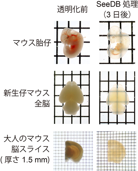
Using fructose found in honey and fruit, along with some water and dashes of other ingredients, researchers created “SeeDB,” short for See Deep Brain, which turns cells transparent. More importantly, it does not cause serious damage to the cells or cause swelling. Basically, it leaves the cells pretty much as they are—but transparent! It takes about three days for the reagent to work. Afterwards, cells look like this:
▼From top to bottom: Mouse embryo, neonatal mouse, adult mouse brain
- Why they did it
For many of us, this probably seems like a big “so what?” It’s not really effective for invisibility (unless you’re only 6 mm thick), so why would we want this? Well, while the average person probably doesn’t have access to all that fancy microscopy equipment, transparency of cells helps out researchers who DO have access to it.
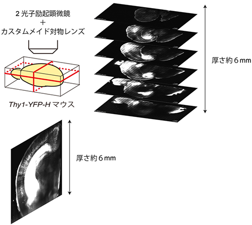
▼Video of 6mm of mouse’s brain, using continuous optical tomography
▼Video of a 3-D cross section of 6mm of a mouse’s brain, taken with two-photon excitation microscopy
Apparently, and I’m going to have to take their word on this, current microscopy only allows scientists to see about 100 micrometers into living cells. Obviously, this isn’t going to let you really “peer into the mind” of anything. By making cells transparent, they’re able to get images of up to 6 mm! Which is an improvement of 600 percent in case you’ve forgotten how many micrometers are in a millimeter. (It’s 1,000, according to the Internet. Because I had no idea.)
▼Two-photon excitation microscopy with a custom made lenses and a cross section of the 6 mm scanned
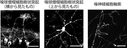
Using this technique, scientists are able to capture images of brains in action, helping map out neural path ways and letting them take closer looks at stuff like the olfactory bulb. Who knows what this information could lead to! Cologne that doesn’t smell like giraffe farts would be nice…
- What’s next
This isn’t just for brains though. The scientists were even able to turn a mouse embryo almost completely invisible (as you saw above). I assume that this is for research purposes, since it would allow them to inject tracers and watch directly how the body develops, and not for more nefarious designs. Like training invisible mouse ninjas. (Or maybe both!?)
▼Left image is of the olfactory bulb mitral cell dendrite from the side, middle image is also of the olfactory bulb mitral cell dendrite from the top, and the right image is of olfactory nerve axons.
But there’s still a lot of work to be done with the old noggin’. The researchers who developed SeeDB believe that this will be useful for analyzing and developing connectomes (basically brain maps) in addition to helping us better understand developmental biology and developing “3-D biology.”
Okay, that last one sounds like something James Cameron dreamt up while sea sick and puking into the ocean.
Source: Riken Center for Developmental Biology (detailed paper, press release)
 Japanese researchers learn how to grow hair follicles, and probably new hair
 Japanese researchers create chickens that lay eggs containing valuable medicine
 Wasabi found to promote hair growth 3 times faster than minoxidil
 Robot finger covered in living skin developed by University of Tokyo
 Anime featuring anthropomorphized blood cells confirmed, actually looks cooler than it sounds
 McDonald’s new Happy Meals offer up cute and practical Sanrio lifestyle goods
 All-you-can-drink Starbucks and amazing views part of Tokyo’s new 170 meter-high sky lounge
 More foreign tourists than ever before in history visited Japan last month
 Starbucks reopens at Shibuya Scramble Crossing with new look and design concept
 Beautiful Sailor Moon manhole cover coasters being given out for free by Tokyo tourist center
 Studio Ghibli glasses cases let anime characters keep an eye on your spectacles
 Tokyo luxury hotel offers month-long stays with free breakfasts, might be cheaper than apartment
 Mister Donut ready to make hojicha dreams come true in latest collab with Kyoto tea merchant
 Hamster abandoned at Tokyo ramen restaurant gets new home
 Inflation making penguins in Japan unhappy with aquarium’s switch to cheaper fish
 Disney princesses get official manga makeovers for Manga Princess Cafe opening in Tokyo
 Beautiful new Final Fantasy T-shirt collection on the way from Uniqlo【Photos】
 Is the new Shinkansen Train Desk ticket worth it?
 Foreign English teachers in Japan pick their favorite Japanese-language phrases【Survey】
 Japanese convenience store packs a whole bento into an onigiri rice ball
 We try out “Chan Ramen”, an underground type of ramen popular in the ramen community
 Studio Ghibli releases Kiki’s Delivery Service chocolate cake pouches in Japan
 Japan’s bone-breaking and record-breaking roller coaster is permanently shutting down
 New definition of “Japanese whiskey” goes into effect to prevent fakes from fooling overseas buyers
 Our Japanese reporter visits Costco in the U.S., finds super American and very Japanese things
 Studio Ghibli unveils Mother’s Day gift set that captures the love in My Neighbour Totoro
 Foreign passenger shoves conductor on one of the last full runs for Japan’s Thunderbird train
 Domino’s Japan now sells…pizza ears?
 New Japanese KitKat flavour stars Sanrio characters, including Hello Kitty
 Kyoto creates new for-tourist buses to address overtourism with higher prices, faster rides
 Sales of Japan’s most convenient train ticket/shopping payment cards suspended indefinitely
 Sold-out Studio Ghibli desktop humidifiers are back so Totoro can help you through the dry season
 Japanese government to make first change to romanization spelling rules since the 1950s
 Ghibli founders Toshio Suzuki and Hayao Miyazaki contribute to Japanese whisky Totoro label design
 Doraemon found buried at sea as scene from 1993 anime becomes real life【Photos】
 Tokyo’s most famous Starbucks is closed
 One Piece characters’ nationalities revealed, but fans have mixed opinions
 We asked a Uniqlo employee what four things we should buy and their suggestions didn’t disappoint
 Princesses, fruits, and blacksmiths: Study reveals the 30 most unusual family names in Japan
 Japanese teens develop anti-fart underwear that blocks smell and sound of flatulence
 Cup Noodle makers successfully create lab-grown diced steak with authentic texture
 Thai mother endures more than 30 hours of labor to deliver stillborn baby in the name of science
 Japanese Fitness expert recommends toddlers play catch, climb on jungle gym, sumo wrestle
 New study suggests Japanese people born in late winter at higher risk of suicide
 Pikachu, Hello Kitty Elected 2025 Expo Ambassadors to Osaka
 Anime’s top catchphrases & top Japanese Internet buzzwords of 2014 announced
 Honda readying a Fuel Cell Vehicle for buyers next year, just in time to challenge Toyota
 Tokyo University research team develops world’s first hybrid robot with real working muscles
 Japan introduces a new kind of needleless injection, you won’t believe how it works!
 Photos of “invisible people” wearing frozen jeans take Japanese Twitter by snowstorm 【Photos】
 Is boob-shaped controller a clever gag ad, pie in the sky dream, or the future of gaming? 【Video】
 They have teeth in their stomaches! And 6 other fun facts about cockroaches
 Take a look around Tokyo Game Show 2016 with us!【Photos】
 Research from Tohoku University claims video games impair brain development in children
Leave a Reply